Key Takeaway:
SEM resolves surface detail and elemental composition, but is unable to see inside the part. Micro-CT delivers non-destructive 3D internal imaging, keeping the part intact for further analysis. The two are often used in sequence: CT first to locate the defect, SEM next for surface analysis. This sequencing saves samples and protects the evidence chain in disputed failures. Our in-house 3D CT supports non-destructive failure analysis, with partner labs coordinated for SEM work.
Table of Contents
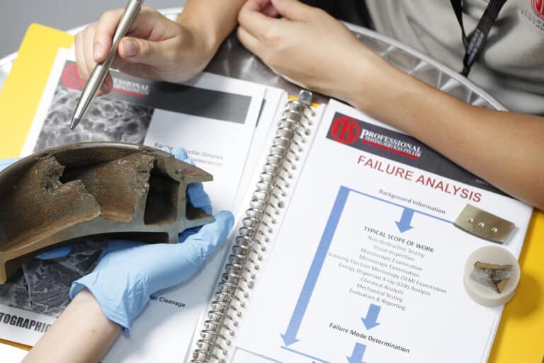
When a part fails, the first instinct is often to send it for SEM imaging. It’s a sensible default as SEM has been the workhorse of failure analysis labs for decades, and for fracture surfaces, it’s still hard to beat. However, SEM only sees the surface, and a lot of failure modes live below it.
This is where micro-CT comes in, being able to change what’s actually visible in an investigation. If you’re unsure how micro CT vs sem imaging line up, this guide breaks down what is the difference between a CT scan and a SEM scan, where each method fits, and how engineers typically use them together when investigating real failures.
What SEM Imaging Does Well
So what is SEM imaging used for? Scanning electron microscopy (SEM) uses an electron beam to produce high-resolution surface images. At its best, SEM delivers nano-scale detail of fracture surfaces, with energy-dispersive X-ray spectroscopy (EDS or EDX) often paired alongside to map elemental composition at the same time.
In metallurgical failure analysis, engineers use SEM to identify fracture modes. Ductile, brittle, fatigue, and intergranular each leave a distinctive surface signature that SEM can resolve clearly. SEM is also the standard for thin films, coatings, and surface contamination.
The hard limit, though, is that SEM is surface-only. If a failure lies inside the part, the sample needs to be sectioned to expose that internal feature. Sectioning is destructive and narrows options once a sample is cut.
What Micro-CT Does Differently
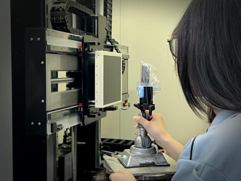
Micro-CT takes a different approach to the same problem. It uses high-resolution 3D X-ray imaging to capture internal structure, with the part staying intact throughout. The process does not involve sectioning, coating, or any destructive measures, allowing for proper non-destructive internal analysis.
Inside that 3D data set, engineers can navigate the internal geometry slice by slice, where voids, cracks, delamination, and assembly defects all show up in their original location. Volumetric measurements can also be taken directly from the scan, and the failed region can be reviewed from every angle without ever touching the part.
Comparing Micro-CT and SEM
Here’s how the two methods stack up across several important factors:
SEM | Micro-CT | |
What it sees | Surface | Internal volume |
Resolution | Nano-scale | Micron-scale |
Sample preparation | Requires sectioning and coating (e.g., sputter coating) | Minimal to no prep required |
Destructive | Usually destructive after sectioning | Non-destructive |
Best suited for | Fracture surface morphology | Internal defect mapping, assembly verification |
When to Choose 2D Digital Radiography
Digital radiography NDT is the right call when:
- Volume is high and weld inspection is the focus, with predictable defect orientation
- Geometries are simple and the defect orientation is known
- Budget matters and flat imaging is sufficient for the defect type
- The project specification mandates digital radiography with predefined acceptance criteria based on established standards
- The work falls into typical zones like oil and gas piping, structural welds, or castings with known critical sections
When Each Method Fits Best
SEM is the right method when:
- The failure is on a fracture surface
- Elemental composition matters (with SEM EDX)
- Surface morphology is the answer
Micro-CT is the right method when:
- The defect is suspected internally
- The part can’t be destroyed before further investigation
- Assembly integrity is in question
In real investigations, the two are often used in sequence. Micro-CT vs SEM failure analysis isn’t always an either-or call, and often micro-CT comes first to locate the defect non-destructively. If surface analysis is then needed at the defect site, targeted sectioning can be done for SEM follow-up.
This sequencing saves samples and protects the evidence chain, which matters a lot in disputed failures or warranty claims.
Why Engineers Lean on Micro-CT for Internal Defects
For certain defect types, micro-CT is the only practical option for:
- Internal voids in castings
- Weld porosity
- BGA solder joint inspection
- Delamination in composites
- Internal cracks in electronics and semiconductor packages
The data also supports work beyond defect analysis. STL model export from CT scan data can be used for reverse engineering reference, which is useful when the original drawings have been lost or when benchmarking competitor parts. For instance, at PTS, we provide STL models (not CAD models) for our 3D reverse engineering services.
For electronics and semiconductor packages, where destructive sampling can destroy the very evidence engineers need, the non-destructive nature of CT is often the deciding factor.
How We Handle Failure Investigations at PTS
At PTS, our laboratory is accredited to ISO/IEC 17025:2017 by SAC-SINGLAS, covering our scheduled mechanical, chemical, metallurgical, and NDT services. Our in-house 3D X-ray computed tomography capability supports non-destructive failure analysis from cell-level electronics to industrial castings.
Engineers can scope investigations with our team to determine the right method or method combination for the failure mode under investigation. Our reports cover defect identification, sizing, and location, with documentation that supports root cause analysis, supplier accountability, and warranty disputes.
For investigations that also require SEM or SEM EDX work, we coordinate with partnering laboratories so the full investigation can be delivered without the need for you to manage multiple labs yourself.
Start Your Failure Investigation With Our Team
In failure analyses, SEM gives the cleanest answer when the question is about a fracture surface. Micro-CT gives the cleanest answer when the question is about what’s happening inside the part. Most serious failure investigations end up using both, in the right sequence, which often saves samples and shortens investigations.
References:
ISO/IEC 17025:2017 – General requirements for the competence of testing and calibration laboratories. Retrieved 22 June 2026, from https://www.iso.org/standard/66912.html
Singapore Accreditation Council (SAC-SINGLAS) – Laboratory Accreditation. Retrieved 22 June 2026, from https://www.sac-accreditation.gov.sg/
Frequently Asked Questions About Micro-CT vs SEM Failure Analysis
A CT scan uses 3D X-ray imaging to capture the internal volume of a part non-destructively, producing slices in any plane. A SEM scan uses an electron beam to produce high-resolution images of the surface only, typically requiring the sample to be sectioned and coated. CT shows the inside; SEM shows the surface.
Neither is universally better; they answer different questions. SEM is better when the failure is on a fracture surface and elemental composition matters. Micro-CT is better when the defect is internal or the part can’t be destroyed. Most thorough failure investigations use both, typically with micro-CT first to locate the defect and SEM second for targeted surface analysis.
PTSPL provides 3D X-ray computed tomography (including micro-CT) in-house at our Singapore laboratory. For SEM and SEM EDX work, we coordinate with partnering laboratories so engineers can have the full failure investigation managed through a single point of contact.
No. Micro-CT is a non-destructive inspection method. The part stays intact through the scan and can be used for further testing, returned to service if appropriate, or preserved as evidence in disputed failures or warranty claims.
CONTACT PTSPL TODAY
Reach out to PTS today for 3D X-ray computed tomography and non-destructive failure analysis at our accredited Singapore laboratory. Our team can scope the right method for your investigation with SEM and SEM EDX coordinated through partner labs where needed, so send us your sample details and we’ll come back with a recommendation. Explore the full range of our Singapore testing services for more.